Culham Research Group
Just another blogs.reading.ac.uk site
Skip to content
  • Home
  • About
  • Advent Botany
    • Advent Botany 2014
    • Advent Botany 2015
    • Advent Botany 2016
    • Advent Botany 2017
    • Advent Botany 2019
    • Taxonomic index to #AdventBotany
  • PhD Students
    • Ahmed Gawhari – PhD 2016
    • Ana Simoes
    • Anas Tawfeeq
    • Azi Jamaludin
    • Donald Zulu
    • Jordan Bilsborrow
    • Maria Christodoulou – PhD 2016
    • Tomos Jones
    • Widad Aljuhani – PhD 2015
    • Kálmán Könyves – PhD 2014
    • Marshall Heap – PhD 2014
    • Ahmed El-Banhawy
    • Hassan Rankou
    • Oli Ellingham
    • Aramide Dolapo Oshingboye – L’Oreal Visiting Fellowship 2014/2015
  • Projects
← How many spores?
Sexual Fungi →

Appendage Morphology

Posted on January 21, 2014 by Oliver Ellingham

Amongst the most important visual, superficial features of the powdery mildew are their appendages. These limb-like features arising from the surface of the, sexual spore containing, chasmothecia are important for latching onto the stems and leaves of their hosts. They vary greatly and can help us to distinguish between tribes and genera.

The ectoparasitic, single conidia producing tribe Erysipheae are a good indication of the diversity of forms within a single tribe: appendages are distinct at their tips (or apices) and can be grouped as uncinate-circinate, dichotomously branched or mycelioid (fig. 1).

Figure 1: Appendages of powdery mildew sexual spores (chasmothecia). The 'uncinate-circinate' appendages (left) are said to be the most basal form from which dichotomously branched appendages (middle) have evolved. The most simple, 'mycelioid' appendages (right), then evolved at least twice from the branched type. Such a series of adaptations shows a simple devolution of complexity in the forms.

Figure 1: Appendages of powdery mildew fruiting bodies (chasmothecia). The ‘uncinate-circinate’ appendages (left) are said to be the most basal form from which dichotomously branched appendages (middle) have evolved. The most simple, ‘mycelioid’ appendages (right), then evolved at least twice from the branched type. Such a series of adaptations shows a simple devolution of complexity in the forms. (From Takamatsu, 2013)

Significant debate has arisen as to the pathway along which these have evolved. As recently as 2012 (Braun and Cook) mycelioid apices were considered to be the most primitive of the three (understandable due to their simplicity). However molecular research, focusing on the rDNA ITS region, has shown a devolution of complexity and as such the curled tips of uncinate-circinate appendages have in fact been around longest

Figure 2: Microscope images showing the characteristic branched apices of Erysipheae section Microsphaera (left) and Erysipheae section Uncinula.

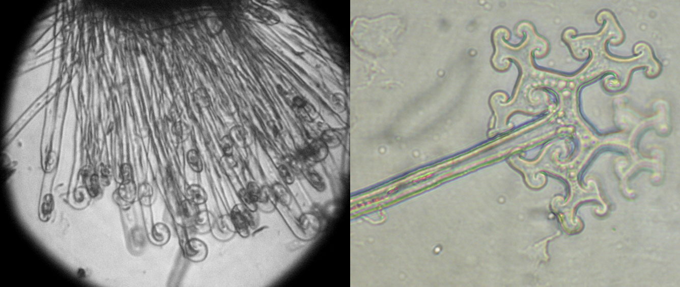
Figure 2: Microscope images accurately showing the characteristic curled apices of Erysipheae section Microsphaera (left) (Image: Steve Clements) and branched apices of Erysipheae section Uncinula (Image: Xerantheum). (From Flickr: The Commons)

Such varied appendages are closely connected to their hosts and overwintering strategies (Takamatsu, 2004). The complex curled and branched apices are required by spores parasitising trees and shrubs as the tougher substrates require more substantial anchorage to last the winter. In contrast the simple, mycelioid appendages are found on pathogens associated with herbaceous plants, which are often protected from harsh, windy, wintery conditions by taking refuge in fallen leaf material and  buds.

References

Braun U, Cook RTA (2012) Taxonomic manual of the Erysiphales (powdery mildews). CBS Biodiversity Series No 11. CBS, Utrecht

Takamatsu, S (2004) Phylogeny and evolution of the powdery mildew fungi (Erysiphales, Ascomycota) inferred from nuclear ribosomal DNA sequences. Mycoscience 45:147–157

Takamatsu, S (2013) Molecular phylogeny reveals phenotypic evolution of powdery mildews (Erysiphales, Ascomycota). Journal of General Plant Pathology (2013): 1-9.

Share this:

  • Facebook
  • Twitter
  • LinkedIn
  • Email
  • Tumblr
  • Pinterest
  • Pocket
  • Print
  • More
  • Reddit
  • Google

Like this:

Like Loading...

About Oliver Ellingham

PhD student at the University of Reading. Working on ID techniques of powdery mildew Fungi. Interested in mycology, plant pathology and arboriculture.
View all posts by Oliver Ellingham →
This entry was posted in PhD research, Public Engagement with Science, RHS research and tagged Appendages, Ascomycota, Erysiphales, Fungal identification, Fungi, morphology, Oliver Ellingham, Pathology, Phylogeny, Plant Pathology, Powdery Mildew, RHS, Royal Horticultural Society, taxonomy. Bookmark the permalink.
← How many spores?
Sexual Fungi →
Logging In...

Leave a Reply to Where it all began… | Culham Research Group Cancel reply

This site uses Akismet to reduce spam. Learn how your comment data is processed.

  • 4 Replies
  • 2 Comments
  • 0 Tweets
  • 0 Facebook
  • 2 Pingbacks
Last reply was December 17, 2015
  1. Fay Newbery
    View January 27, 2014

    I’m confused.
    You are describing appendages of spores but showing the appendages on chasmothecia.
    I thought the sexual spores of a powdery mildew were formed in asci within the chasmothecia.
    Do the spores themselves have appendages? or only the fruiting body – the chasmothecia?

    Reply
  2. Oliver Ellingham
    View January 28, 2014

    Fay, sorry for confusing you, and thanks for pointing that out.
    You are right, of course.
    The appendages “arise from the outer peridial cells of the chasmothecia”.
    The sexual spores within the asci, within the fruiting body, do not have appendages.

    Reply
  3. Where it all began… | Culham Research Group
    View January 29, 2014

    […] stage (teleomorph), relatively large chasmothecia, containing many eight-spored asci, with many uncinate-circinate apiced appendages. The asexual stage (anamorph) produced conidia in chains (Euoidium-type) and […]

    Reply
  4. Powdery mildew morphological glossary | Culham Research Group
    View December 17, 2015

    […] Appendage shape (Fig. 4): Appendages of the chasmothecia enable the structure to adhere to various plant surfaces such as leaves, stems, and bark. Viewing these beautiful structures can help to identify a powdery mildew to genus. […]

    Reply
  • Recent Posts

    • A personal reflection on Wild about Weeds – author Jack Wallington
    • Community fungal DNA workshop
    • #AdventBotany 2019 Day 6: Deck the Halls with Boughs of Holly…
    • Final call: help identify potentially invasive plants
    • #AdventBotany 2019 Day 2: Death by chocolate
  • Recent Comments

    • Specimens, Specimens: Uses | Herbarium World on Brief notes on some Percival Wheats
    • Tendrils: 151218 – The Unconventional Gardener on Advent Botany 2015 – Day 15: Mahleb
    • Tendrils: 151218 – The Unconventional Gardener on Advent Botany 2015 – Day 17: Sgan t’sek
    • Tomos Jones on A personal reflection on Wild about Weeds – author Jack Wallington
    • Future Invaders in your Garden? – The Gardening Zoologist on Future invaders at RHS Chelsea
  • Categories

    • Advent
    • Art
    • Catalogue of Life
    • Collectors
    • e-learning
    • Hawkins Research Group
    • Herbarium RNG
    • i4Life
    • MSc Plant Diversity
    • PhD research
    • Public Engagement with Science
    • RHS research
    • Type
    • Uncategorized
    • Welcome
  • Meta

    • Register
    • Log in
    • Entries RSS
    • Comments RSS
    • WordPress.org
    • RSS - Posts
    • RSS - Comments
  • Subscribe to Blog via Email

    Enter your email address to subscribe to this blog and receive notifications of new posts by email.

  • Privacy & Cookies: This site uses cookies. By continuing to use this website, you agree to their use.
    To find out more, including how to control cookies, see here: Cookie Policy
Culham Research Group
Proudly powered by WordPress.
loading Cancel
Post was not sent - check your email addresses!
Email check failed, please try again
Sorry, your blog cannot share posts by email.
%d bloggers like this: