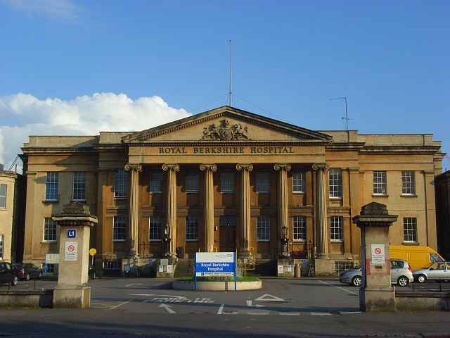
Figure 1. The Hospital, for most people, a dreaded place. For us, a place of revelation
This is the sequel to the post about “Guns” where we talk a little bit about where palaeoecology leads us when we follow our quest for investigating the past using lake/bog sediments.
An unusual patient
If you are worried about the consequences of working in palaeoecology, I can assure you that my recent visit to A&E was not because of an accident (and was not an accident either). This was a very well planned visit to the hospital to become more familiar with my beloved Brazilian patients, my bog cores.
Having taken over 110 Russian cores from numerous bogs across southern Brazil, between Frank Mayle and I (check out the Je Landscapes Project website to know more), I needed a quick non-destructive technique to visualize the internal structures of the cores so I could select the best ones for the project. I therefore decided to take x-rays of these half-metre cores. This relatively low-cost technique allows us to identify any key lithological changes through the core which are not apparent to the naked eye. The differences in density are seen in the resulting images as shades of light and dark. The lighter the colour of the image, the denser the sediment is. That is why features such as clastic material and tephra layers appear light, in comparison with organic peats which are usually dark.
So I arrived at the hospital with two oversized suitcases (Figure 2), completely filled with sediment cores (I am glad to say that I didn’t have to rush there). Carrying the cores this way allowed me to navigate easily through the labyrinthine hospital and get to the subterranean x-ray room.

Figure 2. Just arrived at the Hospital
The X-ray manager was very helpful, and we worked together as a great team, with me unwrapping the cores and placing them on the plate, taking notes and hiding behind the x-ray shield; while he was pressing the button and inputting the information into the computer (Figure 3). It was a relief to be on the other side of that shield.

Figure 3a. Our view to the patient. Behind the shield
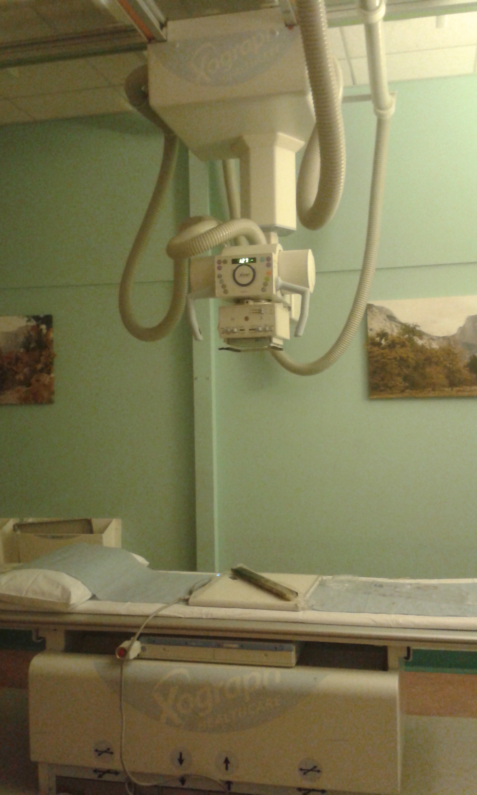
Figure 3b. Not sick looking, one of the bog cores being analyised
The cores just about fitted on the x-ray plate (in diagonal) (Figure 4). I am glad I checked that beforehand! If you are considering taking x-rays of your cores, its important to call the hospital beforehand to make sure they have the plate size you need. It seems that most of the x-ray facilities only have the 23cm length plate, which is no where near large enough for a half-metre Russian core.

Figure 4. X-ray of one of the bog cores
I am pleased with the results. The images allow me to distinguish internal structures and therefore enable me to select the best cores (some cores look fine from the outside, but with the x-rays I can see some small gaps in the sediment). On the other hand, the lithological changes revealed by the x-rays enable me to cross-correlate overlapping and duplicate cores. A particularly useful further step is to undertake grey scale analysis of the x-ray image, which can reveal even greater detail (Figure 5), especially when correlating with XRF and magnetic susceptibility results.
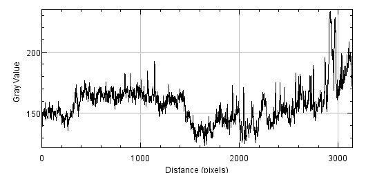
Figure 5. Grey scale analysis performed with ImageJ in one of the cores
This was my first experience of undertaking x-ray analysis of bog cores, and I must say that I am very pleased with how useful this technique has proven to be.